Resources Archives - Peak Health & Performance

Plantar Fasciitis – What? Why? FIX ME!

By: Dr. Claire Wells, DC
Chiropractor, Peak Health Elbow, Calgary, Alberta.

If you’ve ever had pain on the bottom of your heel, you’ve probably been told (or with the help of Dr. Google you’ve told yourself) that you have plantar fasciitis. And you could be right! In this post I’m going to break down the basics of this condition, and what we can do about it. Let’s dive in!

Anatomy Overview

Plantar-Fascisitis

When there is pathology in the plantar fascia, we are specifically referring to a section called the “aponeurosis.” Almost always, there is degeneration of this tissue, as opposed to inflammation. This means the term “fasciitis” is not very accurate (“-itis” indicating inflammation), and “fasciopathy” is more appropriate (“-opathy” indicating an injury or problem with a tissue).

The plantar aponeurosis originates from the calcaneal tubercle (a bump on the bottom of the heel bone) and extends to the forefoot. In the forefoot, it divides into five separate bundles, which attach to the closest part of the toes. The plantar aponeurosis is not identical to a tendon or a ligament – it’s somewhere in-between, and is comprised largely of type 1 collagen. (1)

Fascial Function

The plantar aponeurosis supports the bottom of the foot, especially the medial arch. It plays an important role in turning the foot into a rigid lever during the push-off (“propulsion”) phase of walking and running.(2) But it doesn’t work alone! Other structures support the medial arch as well, including muscles. Strength of these muscles is critical for foot stability, and for preventing the aponeurosis from being over-stressed. Eccentric strength, which is when a muscle is lengthened under load, is especially important, since walking and running involve a lot of joint deceleration. 

When walking or running, the aponeurosis and the associated muscles become tensioned when the big toe extends. This is called the ‘windlass mechanism’ and is critical for efficient movement.(2) Ensuring that we have good range of motion and strength of the big toe is important for healthy plantar fascia. 

So what is plantar fasciopathy?

The problem is excessive biomechanical strain at the insertion of the aponeurosis on the heel bone. When the demands on the tissue exceeds the tissus ability to heal, we get tissue breakdown, or degeneration, and sometimes pain. Inflammation does not typically occur. 

What are the risk factors for developing plantar fasciopathy?

The following factors can be associated with plantar fasciopathy(3, 4). The bold ones are factors that are modifiable (which is great news!). It’s important to realize that if any of these apply to you, it does not mean that you will get plantar fasciopathy, nor does it mean that you can’t do anything about it if you do get it.

  • BMI >27
  • Excessive running or a recent significant increase in weight-bearing activity
  • Intrinsic foot muscle and calf muscle tightness
  • Reduced ankle dorsiflexion range of motion, and/or excessive plantarflexion range of motion
  • Weak calf muscles 
  • Sedentary lifestyle
  • Metabolic conditions such as Type 2 Diabetes
  • Acute increase in loading/exercise
  • Leg length discrepancy
  • Femoral or tibial torsion (This means an excessive structural twisting of the thigh or shin bone; this is something that we cannot change. But having this anatomical variation does not mean you are doomed!) 
  • Occupations that require prolonged periods of walking or standing
  • High arches
  • Low/flat arches

How Do I know if I might have plantar fasciopathy?

Classic signs of plantar fasciopathy include(3, 4):

  • Pain located at the bottom of the heel, or where your heel and arch intersect
  • Usually affects only one foot
  • Worse first thing in the morning, or with the first steps after being prolonged rest (especially if your ankle has been in plantar flexion, aka pointed downwards). It may also be worse in the evening.
  • Pain is sharp or stabbing, and does not radiate
  • No tingling or pins and needles

Do I need to get diagnostic imaging?

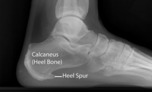
X-rays can’t help us, because this is not a bone problem. Ultrasound and MRI can show the density and thickness of the fascia, but we don’t need that information to diagnose the condition or predict prognosis. So, diagnostic imaging is not necessary unless other injuries like a fascial rupture or a calcaneal stress fracture are suspected.(4)

Differential Diagnoses: What else could my plantar heel pain be?

“Plantar” refers to the bottom surface of the foot. Not all pain on the bottom of the heel is plantar fasciopathy! Other causes of pain in this region can be misdiagnosed as plantar fasciopathy, and thus may end up being treated ineffectively. Peripheral nerve entrapment is a big one. This is when one of the nerves that supplies the bottom of the foot is compressed, or otherwise biomechanically irritated. If this is the cause, you might only get pain, but you might also get numbness, pins and needles, tingling, or burning or electric-like pain. You may also feel worse during or after stretching, or rolling out the bottom of the foot. Nerve pain could also be originating from the low back, so it’s important to rule out the spine as the culprit.
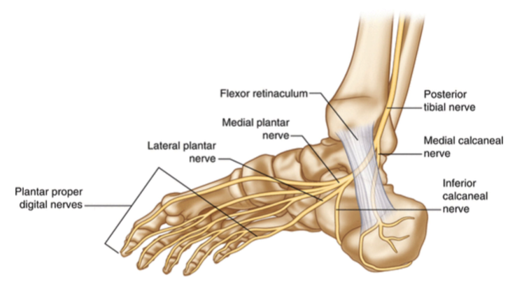
Sometimes you can get plantar heel pain from muscle tightness or trigger points. Other less common conditions that can cause pain in this area include tibialis posterior tendon dysfunction, calcaneal stress fracture, insertional achilles tendinopathy, Sever’s apophysitis, calcaneal fat pad atrophy, and inflammatory arthropathy.

If I have plantar fasciopathy, what can I do about it?

Ensuring that you see a practitioner who can assess your specific case and prescribe an individualized exercise program for your mobility and stability needs is an important step in tackling your pain. 

You may need initial activity modification to reduce pain, but gradual overload of the tissue is required to promote healing and build up capacity for loading. Being out of pain does not equal being ready to immediately dive right back into high activity levels.

Addressing factors that may have contributed to this in the first place is important. Do you have good foot stability and control? If not, you might rely too much on passive structures like the plantar aponeurosis. Do we have good trunk and lower limb stability and control? If not, the whole posterior chain may over-work and tense up, which may cause nerve or muscle tension as far down as the calf and foot. 

Treatment Options for Plantar Fasciopathy

Usually for long-term success, we need to work on intrinsic foot muscle strength, and big toe mobility and strength. But when it comes to treatment that targets the plantar fascia itself, we have research-based exercise protocols that improve function and reduce pain. Keep in mind that because this is a degenerative condition of connective tissue (which takes longer to heal), you will have to be patient with the recovery process. It’s not going to go away within a few days!

When it comes to pain and function, we have a hierarchy of interventions.

  1. Strengthening the plantar fascia is superior to…
  2. Stretching the plantar fascia, which is in turn superior to…
  3. Stretching the Achilles tendon/calf muscles

Good strengthening requires that we get tension in both the Achilles and the plantar aponeurosis, by activating the windlass mechanism. You’ll recall that the windlass mechanism is when big toe extension tensions the aponeurosis and muscles of the medial arch. We invite the Achilles tendon to the party to further increase fascial tension – this works because these two connective tissue structures are, well, connected! (1)

Where to Start:

Single leg calf raises, with the toes supported in maximal extension is a great place to start.(6) There is a specific loading protocol that research has shown to be effective, but it’s best to see your chiropractor or physiotherapist for an exercise program that is tailored to your specific needs. The studies on this used a 12-week program, which is important to keep in mind. It takes time for the tissue to adapt, even if you start to see improvement in pain before 12 weeks.

Shockwave therapy, laser therapy, orthotics, dry-needling, taping, and other techniques provided by chiropractors and physiotherapists can be helpful for improving pain and function, as adjuncts to exercise.

What about injections?

A systematic review and meta-analysis found no high-quality evidence to show that corticosteroid injections are better than other forms of treatment for improving pain and function in the short, medium, or long-term.(9) They have similar outcomes to placebo injections (which means that a real corticosteroid injection was not better than a fake one). They also result in total rupture of the fascia in 2-10% of cases. 

A systematic review and meta-analysis of platelet-rich plasma (PRP) vs. corticosteroid injections found that PRP is better at reducing pain, however, the quality of the studies was generally low and the injection protocols were highly variable, so these results must be viewed with caution.(10)

Overall, I recommend trying conservative treatment first, such as chiropractic or physiotherapy. PRP may be something to consider for severe and/or chronic, unremitting pain. 

Take-home points:

  • Plantar heel pain is not always plantar fasciopathy. Get assessed to make sure

  • The plantar aponeurosis needs help from the muscles of the foot, and good mobility of the big toe, in order to function well

  • Treatment may need to address more than just the foot itself

  • Strengthening is the best treatment! This tissue requires intentional loading to recover properly 

References:

  1. Stecco C, Corradin M, Macchi V, et al. Plantar fascia anatomy and its relationship with Achilles tendon and paratenon. J Anat. 2013;223(6):665-676. doi:10.1111/joa.12111


  1. Wearing SC, Smeathers JE, Urry SR, Hennig EM, Hills AP. The pathomechanics of plantar fasciitis. Sports Med. 2006;36(7):585-611. doi: 10.2165/00007256-200636070-00004.


  1. Trojian T, Tucker AK. Plantar Fasciitis. Am Fam Physician. 2019 Jun 15;99(12):744-750. PMID: 31194492.


  1. Monteagudo M, de Albornoz PM, Gutierrez B, Tabuenca J, Álvarez I. Plantar fasciopathy: A current concepts review. EFORT Open Rev. 2018 Aug 29;3(8):485-493. doi: 10.1302/2058-5241.3.170080. 


  1. Menz HB, Thomas MJ, Marshall M, Rathod-Mistry T, Hall A, Chesterton LS, Peat GM, Roddy E. Coexistence of plantar calcaneal spurs and plantar fascial thickening in individuals with plantar heel pain. Rheumatology (Oxford). 2019 Feb 1;58(2):237-245. doi: 10.1093/rheumatology/key266. PMID: 30204912; PMCID: PMC6519610.


  1. Rathleff, M. et al. “High‐load strength training improves outcome in patients with plantar fasciitis: A randomized controlled trial with 12‐month follow‐up.” Scandinavian Journal of Medicine & Science in Sports 25 (2015)


  1. Riel H, Jensen MB, Olesen JL, Vicenzino B, Rathleff MS. Self-dosed and pre-determined progressive heavy-slow resistance training have similar effects in people with plantar fasciopathy: a randomised trial. J Physiother. 2019 Jul;65(3):144-151. doi: 10.1016/j.jphys.2019.05.011. Epub 2019 Jun 13.


  1. Salvioli S, Guidi M, Marcotulli G. The effectiveness of conservative, non-pharmacological treatment, of plantar heel pain: A systematic review with meta-analysis. Foot (Edinb). 2017 Dec;33:57-67. doi: 10.1016/j.foot.2017.05.004. Epub 2017 Jun 15. PMID: 29126045.


  1. Whittaker GA, Munteanu SE, Menz HB, Bonanno DR, Gerrard JM, Landorf KB. Corticosteroid injection for plantar heel pain: a systematic review and meta-analysis. BMC Musculoskelet Disord. 2019 Aug 17;20(1):378. doi: 10.1186/s12891-019-2749-z. PMID: 31421688; PMCID: PMC6698340.


  1. Hohmann E, Tetsworth K, Glatt V. Platelet-Rich Plasma Versus Corticosteroids for the Treatment of Plantar Fasciitis: A Systematic Review and Meta-analysis. Am J Sports Med. 2021 Apr;49(5):1381-1393. doi: 10.1177/0363546520937293. Epub 2020 Aug 21.

Running Myths

By: Dr. Claire Wells, Chiropractor

Running is one of the most accessible forms of exercise, and a great way to enjoy being outside during these remaining weeks of good weather in Calgary. Because so many people run, there is a lot of information that circulates on social media, in marketing campaigns, and even by word of mouth, about what we “should” do when it comes to running. But not all information is good information, so below I bust four common running myths to help you get the most out of the season!

MYTH 1: “You should minimize rotation of the trunk and arms.”

When I started running, I was certain that rotating my body was bad. I was told it was a waste of energy; that my arms should never stray from straight, and my hips should always face perfectly straight forward. I was so proud when I would go for a run and keep my trunk super rigid and stiff.

But wow, was I wrong! As it turns out, running is a rotational sport. Controlled rotation throughout the body is how we propel ourselves forward. Let me explain…
It would be easy to think of the legs as being our primary drivers for moving our body forwards when we run (after all, that’s what it looks like!). But even if you had no legs, you would be able to “walk” by reciprocal rotation of the pelvis (driven by the lumbar spine, AKA the low back) and the shoulders (driven by the thoracic spine AKA the mid and upper back).

[Image1]

“It’s important to realize that trunk, hip, knee, ankle, and foot rotation is normal and necessary to run efficiently. And efficiency means reduced injury risk.”

This is not to say that the legs don’t matter! Even though the trunk is our driving force, the legs play a huge role in running, and they also must rotate. It’s important to realize that trunk, hip, knee, ankle, and foot rotation is normal and necessary to run efficiently, which means reduced injury risk.

Now, this doesn’t mean we should go out and run like a wacky inflatable tube man, flailing our body parts into twisting motions. There is such a thing as unhelpful rotation (if it’s not producing momentum to propel us forward, it’s not helpful), so there is a sweet spot when it comes to how much rotation we produce at each level of the body. And most importantly, if we do not have control of the ranges we use when running, we are setting ourselves up for some problems.

[Image2]

My Advice: 

Be able to resist rotation before you can expect to create it efficiently and be able to control the range of motion available to you. This means doing specific exercises to build up your capacity (and yes, you need to strength train if you are a runner!). It’s best to see a chiropractor or physiotherapist in Calgary who can identify deficiencies specific to you, and prescribe exercises accordingly but here are a few examples:

• Core/trunk: Paloff press, bear plank or high plank shoulder taps, cable chops and lifts

• Hip: single leg Romanian deadlifts, hip airplanes, 90-90 get-downs, rotational cable squats

• Ankle and foot: banded supinations, ankle rocker board, active inversion-eversion

Unlike me when I started running, don’t think you need to keep your shoulders and hips rigidly facing forward when you run. Let your “spinal engine” do its thing!

MYTH 2: “You should forefoot strike to reduce impact on your joints”

You may have heard that landing on your heel is “bad for your joints,” or even just that it’s inefficient. Don’t believe the lies, friends! There is no inherently superior strike pattern during running.

Our bodies are smart, not to mention highly variable in individual anatomy, and your body will self-select the strike pattern that is mechanically efficient for you. Speed of running will also change this. Until you reach a 6:00-6:30 minute-mile pace, heel striking is more efficient for most people. At that pace, heel and midfoot striking are about equally efficient. Most elite marathoners midfoot strike because they are running so fast. Similarly, you’ll notice that sprinters are always landing on the forefoot, out of necessity, because of their speed.

Slow running is more efficient with heel striking. This means runners who naturally heel strike should not try to intentionally forefoot strike. Doing so may overload the gastrocnemius (not as well designed to absorb force) and can lead to posterior calf (and Achilles tendon) injury.

[Image3]

When we change our strike pattern, we change what structures are predominantly absorbing the force, but we do not change how much force the body needs to absorb. [4]

• Forefoot strike: ankles and calves

• Midfoot strike: foot arches and tibialis posterior

• Heel strike: knees and tibialis anterior

So, good news: There is no difference in injury rate between forefoot and rearfoot striking, and there is no association between foot strike pattern and performance. [2, 3, 7] Thus, no need to force yourself to change what you do.

Note: I am not advocating that everyone heel strike, just recommending that you don’t try to intentionally change your heel strike pattern, unless you have significant knee pain, for example. But if that’s the case, see a chiropractor or physiotherapist!

My Advice:

What most research seems to support is that what we should care about, more than foot strike, is the angle of the tibia (the shin bone) when we make contact with the ground. We want to see at most a 7-degree angle in front of vertical. Vertical, as in the shin being perpendicular to the ground (or straight up from the foot to the knee), is ideal. Not sure if you do this? Try having someone film you running, take some screenshots, and then use a free app like Technique or Hudl to draw angles on the photo.

[Image4]

MYTH 3: “You should wear a supportive shoe to minimize risk of injury”

Ultra-cushioned and motion-control shoes are becoming more popular because marketing campaigns have us believing that they can better attenuate shock, and thus reduce injuries. But…do they? Let’s take a look at some research…

[Image5]

“Extra cushioning interferes with proprioception, and therefore slows reaction time. This means our ability to stabilize and control our lower extremity is compromised, and you may get injuries higher up the chain.”

One study found that in the ultra-cushioned shoes, the timing was off for normal joint motion between the calcaneus (heel bone) and tibia (shin bone). This resulted in increased rotational stress on the knee, which may contribute to knee injury.

→ Super-cushioned shoes are unlikely to benefit your mechanics in a way that is protective or advantageous. [8]

One study followed 952 runners for a year. They all ran in a neutral running shoe, regardless of their foot type, and there was no significant difference in the risk of sustaining an injury for any foot type. This makes sense because foot type is NOT consistently associated with injury rates in research. [9]

In a study that followed runners training for a half-marathon, they found that those wearing the motion control shoes had significantly greater pain, regardless of foot type.

→ They concluded that picking shoes based on stability categories does not reduce the risk of running pain. [11]

This might go against what seems intuitive, which is that sloppy feet need to be controlled. But, excessive cushioning decreases the foot’s ability to get sensory information and send it to the brain. Reduced sensory input to the brain equals reduced quality of motor output (we can’t react to what we don’t know). We need constant feedback of impact forces and foot position to control ourselves. [10]

For this reason, it makes sense that wearing a highly motion-controlled or cushioned shoe could actually be WORSE for someone with feet that pronate too early or for too long. Extra cushioning interferes with proprioception, and therefore slows reaction time. This means our ability to stabilize and control our lower extremity is compromised, and you may get injuries higher up the chain.

My Advice:

The take home here is that you don’t need a specific type of shoe for a specific type of foot. Just buy shoes that feel COMFORTABLE. Your toes should be able to splay within the shoe, so if you can get a wide toe box on your shoes, even better.

MYTH 4: “You should stretch to prevent injuries”

NO studies have shown that “acute stretching,” aka stretching right before a run, improves running performance. It might actually decrease it! And long-term stretching doesn’t seem to make for better running, either.

Here are some brief examples based on research: [12]

• Stiffer thigh and calf muscles equals better force transfer between the deceleration and push-off phases.

• Less flexible hip and calf means less muscular effort needed to stabilize during foot strike

• 10 weeks of stretching resulted in no improvement in running efficiency, and no reduction in injury incidence. In this case, the stretching was even done separately from running.

• Gene COL5A1 is associated with inflexibility, and is found in elite endurance (runners with this gene had significantly higher running efficiency than others in one study)

• Stretching has no effect on chronic injury prevalence in runners

[Image6]

Elastic energy is what makes running efficient. The changes that occur from stretching reduce our ability to store and release elastic energy. So, we need more muscular effort to stabilize our joints.

High muscular effort = more oxygen needed = it takes more work to run!

This applies to stretching right before a run, and also long-term. Now, this doesn’t mean nobody should stretch. It really depends on how much range of motion you already have (and control of that motion, like I mentioned in Myth 1). There is a sweet spot, where you have enough range of motion to allow for the required movement, but not so much that we sacrifice elastic energy.

Another interesting thing to note is that there is also no evidence that stretching has the ability to reduce the severity or duration of delayed onset muscle soreness (DOMS). So, for most of us, stretching is not going to prevent injuries, increase performance, or reduce soreness. But sometimes it feels good to do!

My Advice: 

Go ahead and stretch if it feels good for you, but I’d recommend against doing a ton of it, and definitely not before a run, in the name of mechanical efficiency. Your time could be better spent on other activities to support your running goals.

Take-home points:

1. Let your body rotate while running

2. Don’t force a specific foot strike

3. When choosing shoes, just go with what feels comfortable

4. Stretching does not make you faster or less likely to be injured

These are general principles that will apply to most people, but if you are a runner and you’re experiencing pain or have questions about what running factors are specific to you, it’s best to get assessed by a chiropractor or physiotherapist. Our teams at Peak Health Elbow and Peak Health Marda will get you back to that runner’s high and you can book online here.

References:

1. Gracovetsky, Serge. (1997). Linking the spinal engine with the legs: a theory of human gait. Movement, Stability and Low Back Pain – The Essential Role of the Pelvis.

https://cdn2.hubspot.net/hub/52884/file-5411457.pdf/2012%20NCAA%20Indoor%20Track%20Championships

2. Kasmer ME, Liu XC, Roberts KG, Valadao JM. The Relationship of Foot Strike Pattern, Shoe Type, and Performance in a 50-km Trail Race. J Strength Cond Res. 2016 Jun;30(6):1633-7. doi: 10.1519/JSC.0b013e3182a20ed4. PMID: 23860289.

3. Kasmer ME, Liu XC, Roberts KG, Valadao JM. Foot-strike pattern and performance in a marathon. Int J Sports Physiol Perform. 2013 May;8(3):286-92. doi: 10.1123/ijspp.8.3.286. Epub 2012 Sep 19. PMID: 23006790; PMCID: PMC4801105.

4. Kleindienst F, Campe S, Graf E, et al. Differences between fore- and rearfoot strike running patterns based on kinetics and kinematics. XXV ISBS Symposium 2007, Ouro Preto, Brazil.

5. Hasselman C, Best T, Seaber A, et al. A threshold and continuum of injury during active stretch of rabbit skeletal muscle. Am J Sports Med. 1995;23:65-70

6. Cunningham C, Shilling N, Anders C, et al. The influence of foot posture on the cost of transport in humans. J Experimental Biol. 2010;213:790-797.

7. Miller R, Russell E, Gruber A, et al. Foot-strike pattern selection to minimize muscle energy expenditure during running: a computer simulation study. Annual meeting of American Society of Biomechanics in State College. PA 2009.

8. Brianne Borgia, Julia Freedman Silvernail & James Becker (2020): Joint coordination when running in minimalist, neutral, and ultra-cushioning shoes, Journal of Sports Sciences, DOI: 10.1080/02640414.2020.1736245.

9. Nielsen RO, Buist I, Parner ET, Nohr EA, Sørensen H, Lind M, Rasmussen S. Foot pronation is not associated with increased injury risk in novice runners wearing a neutral shoe: a 1-year prospective cohort study. Br J Sports Med. 2014 Mar;48(6):440-7. doi: 10.1136/bjsports-2013-092202. Epub 2013 Jun 13. PMID: 23766439.

10. Richards, C E; Magin, P J; Callister, R (2009). Is your prescription of distance running shoes evidence-based?. , 43(3), 159–162. doi:10.1136/bjsm.2008.046680.

11. Ryan MB, Valiant GA, McDonald K, et alThe effect of three different levels of footwear stability on pain outcomes in women runners: a randomised control trialBritish Journal of Sports Medicine 2011;45:715-721.

12. Claire Baxter, Lars R. Mc Naughton, Andy Sparks, Lynda Norton & David

Bentley (2016): Impact of stretching on the performance and injury risk of long-distance

runners, Research in Sports Medicine, DOI: 10.1080/15438627.2016.1258640

Media References:

• [Image1] https://erikdalton.com/blog/legs-really-necessary/

• [Image2] https://www.tunnelmarathon.com/blogs/news/counter-rotation-exercises-for-runners

• [Image3] https://www.roadrunnersports.com/blog/running-foot-strike/

• [Image4] https://www.nurvv.com/en-us/support/footstrike/

• [Image5] https://supinate.wordpress.com/tag/supination/

• [Image6] https://www.womenshealthmag.com/uk/fitness/running/a706581/best-calf-stretches-for-runners/

What is Mechanical Low Back Pain?

By: Cody Gramlich, Physiotherapist

Most of us have been there before. You go to pick something up off of the floor and there it is. Back pain so severe you feel like you may never be able to walk again. An alternative scenario is pain that won’t stop nagging throughout your workday. You could also be someone who notices a ‘twinge’ every time you attempt a back squat. Regardless of the situation, a LOT of people experience back pain. If you haven’t, you  are one of the lucky ones in the minority. The good news is, with proper understanding and management, it doesn’t have to define you! If any of this sounds familiar to you, the information below will be of benefit. You will gain a better understanding of back pain, how to manage it, and how to prevent it.

What is Mechanical Low Back Pain?

Starting with a very general overview of the anatomy of the lumbar spine (low back), it consists of 5 vertebrae, separated by intervertebral discs. The vertebrae provide structural support, guide movement, and provide protection for your spinal cord and spinal nerves. Your intervertebral discs provide shock absorption from impact activity and bending.  Your lumbar spine also has various ligaments and muscles that contribute to structural support and movement. As you can see in the second image below, there is close interaction between the areas above and below your spine due to the overlap of multiple ligaments and muscles. These muscles help facilitate movement of your low back in 3 different planes:

  • Flexion/extension (forward/backward)
  • Lateral flexion (side bending)
  • Rotation (twisting)

Mechanical low back pain (also referred to as non-specific low back pain) is defined as “low back pain not attributed to recognizable, known specific pathology”3. As the name and definition suggest, you cannot conclusively determine the structure at fault for mechanical low back pain. Many research studies have indicated that even imaging (x-ray, CT scan, MRI) does not always correlate with clinical findings when it comes to low back pain. To hammer this point home, one of my mentors and colleagues used to say, “we could sit down and drink a few pitchers of beer arguing what we think the specific cause of mechanical low back pain is”.

When your low back pain is being assessed, the first step is to rule out any 
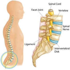
specific/serious causes of your pain. Examples of this include, but are not limited to, radiculopathy (nerve related deficits), cauda equina syndrome, tumor, fracture, infection, and inflammatory disease. If these more specific causes are ruled out, it is then labelled as non-specific mechanical low back pain. You may be asking yourself “how can a physiotherapist help if they cannot identify the cause of my back pain?”. The goal of assessment/treatment with a physiotherapist will be to manage the symptoms and identify contributing factors that may have led to the development of it in the first place.

Potential contributing factors to the development of your low back pain3,4:

– History of trauma

– Strain or overuse

– Postural dysfunction

– Dysfunction above and below the low back (mid back, pelvic girdle, hips)

– Core weakness or muscle imbalances

– Psychological factors (ex. fear of movement, depression)

– Work environment

– Pregnancy

Since mechanical low back pain is so common, there has been a significant amount of research surrounding it, producing clinical practice guidelines (best practices) for diagnosis and management. A stratified approach is now most commonly suggested. The recommendations for management of your low back pain will differ depending on what group or subgroup you are categorized into. These groups/subgroups are usually based on chronicity of your back pain (acute, sub-acute, chronic) and presence of external factors that may contribute to it (psychological, work environment, etc.)1,3,4,6.

Why is it Important to Understand Mechanical Low Back Pain?

Low back pain affects 60-80% of individuals at some point in their lifetime3. Of these 
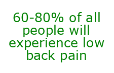
people, over 90% that present to a primary care practitioner have non-specific mechanical low back pain4. Back pain is also a significant economic burden to society. It is reported that the cost of care for low back pain is $50 billion annually in the United States4. Up to 23% of the world’s adults suffer from chronic low back pain (lasting longer than 3 months), which requires significant health care costs3. Without diving too deep into politics, in Canada’s public health care system and considering the significant costs associated with back pain, you could certainly argue it affects us all.

It is important to note that low back pain tends to be a self-limiting condition. Half of individuals will recover from it in 2 weeks without treatment, and the majority of individuals will recover in 1-4 months without treatment4. Although this is true, the recurrence rate of low back pain is high. 60% of people are likely to experience another episode of low back pain within 3-6 months4. These statistics indicate why it is important to see a rehab professional. First, some guidance will give you a higher likelihood of recovering in a shorter time frame. Second, education on your back pain will allow you to take preventative steps to decrease the likelihood of recurrent episodes.

As mentioned earlier, current guidelines take a stratified approach to diagnosing and treating mechanical low back pain. Depending on where you are categorized, the 
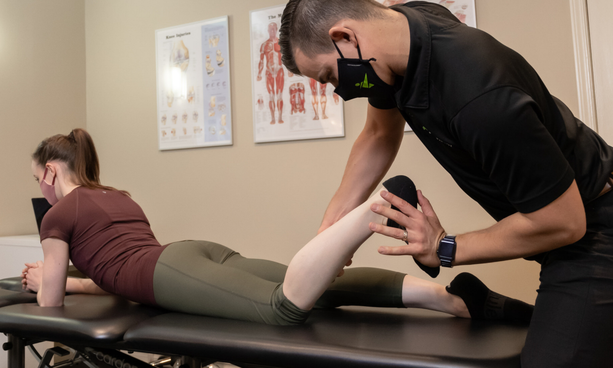
treatment may differ slightly. However, within each category, a few common themes exist. First, one of the most important aspects of care is providing reassurance and education. The better you understand your low back pain, the better you will be equipped physically and psychologically to deal with the pain. Second, exercise therapy and maintaining an active lifestyle is a key component to preventing and managing it. When it comes to mechanical low back pain, as long as a primary care practitioner has ruled out red flags, it is safe to move, and encouraged to help recovery. Finally, a multidisciplinary approach to low back pain management is recommended. This could include (but is not limited to) pharmaceuticals, psychology, conservative care, and self management1,2,4,5.

Some Risk Factors for Developing Mechanical Low Back Pain4:

– Standing or walking > 2 hours per day

– Frequent moving or lifting > 25lbs

– Increased driving time (occupational)

– Limping or altered gait

– Obesity

– Psychosocial factors (income level, stress level, poor relationships at work)

– Prior low back pain

– Posture

– Poor muscular endurance (low back and core)

How Do I Know If I Have Mechanical Low Back Pain?

– Pain, ache, or stiffness in the lumbosacral region (small of your back)

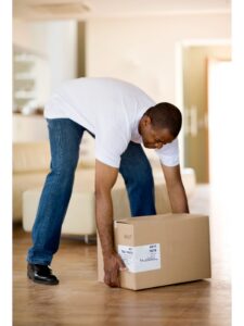
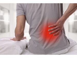
– Pain may radiate into your buttocks or upper leg (more often one sided)

– Pain may come on due to a specific event (bend, lift) or insidiously (gradual, no cause)

– Pain will increase or decrease with positional changes, certain movements, or lifting

– NO signs of Red Flags (i.e. significant trauma, unexplained weight loss, widespread neurological issues)

– NO known specific pathology (i.e. fracture, infection)

*Remember that these are the most common symptoms that would indicate you may have mechanical low back pain. It may present with different signs/symptoms depending on the individual. If this sounds like you, reach out to a Peak Health physiotherapist or another health care practitioner for a thorough assessment to determine the cause of your specific symptoms.

3 Strategies to Help Manage Your Mechanical Low Back Pain Symptoms

1. Keep it Moving

*Movement and activity are a key component to recovery and prevention of mechanical low back pain. Pick an activity you like to do (yoga, walking, cycling) and do it within the limits of your pain levels. You may have to modify the activity in the short term, but the movement will be beneficial for your body.

2. Low Back Mobility

*Working on mobility in your lumbar spine will help decrease pain levels and encourage more efficient movement in the long term. Try 3 sets of 10-15 repetitions.

3. Back/Core Strengthening and Motor Control

*Strength and coordination of the back, hip, and core muscles will allow you to get back to normal activity sooner and prevent episodes of back pain in the future. Try 3 sets of 8-10 repetitions on each side.

FAQ

How Do I Relieve My Low Back Pain?

It is best that you take an active approach or incorporate movement to help alleviate your back pain. Another suggestion includes using heat or ice to help decrease pain or muscle spasm. Clinical practice guidelines also recommend short term use of over-the-counter anti-inflammatory medications. However, it is best to consult a physician or pharmacist prior to use to ensure medication is safe and effective for you.

When Should I Be Worried About My Low Back Pain?

If your low back pain is non-specific mechanical low back pain, there is no need to be worried, as your pain will likely resolve with multimodal treatment. Mechanical low back pain is self-limiting, and it is usually safe to resume activity as tolerated. However, it is best to have it assessed by a physiotherapist or another health care professional in order to rule out red flags or specific causes of your pain that may require more immediate attention.

How Should I Sleep With Low Back Pain?

Most comfortable sleeping position will vary from person to person and there is no right or wrong answer. You may have to try a few different positions to find out what is best for you. One option to try is sleeping on your back with a pillow supporting your knees. Another option is sleeping on your side with a pillow between your legs. These options can put your pelvis and low back in a position that may relieve some discomfort.

What Comes Next?

Remember, mechanical low back pain is not attributable to any recognizable or specific pathology. It is one of the most common complaints to primary care practitioners and has a large burden on the health care system. Mechanical low back pain can be complex and multifactorial. It usually requires individualized management using a stratified approach. Education, an active lifestyle, and multidisciplinary management will aid in more successful outcomes.

Start by trying some of the strategies listed above and see how it goes! Afterwards, it would benefit you to see a physiotherapist to guide you through treatment, depending on your response.

Feel free to reach out if you have any additional questions on mechanical low back pain, or you can book an appointment online by clicking here.

References:

  1. Almeida, M., Saragiotto, B., Richards, B., & Maher, C. G. (2018). “Primary care management of non-specific low back pain: key messages from recent clinical guidelines”. The Medical Journal of Australia, 208(6), 272–275. doi:10.5694/mja17.01152
  2. Oliveira, C.B., Maher, C.G., Pinto, R.Z. et al. “Clinical practice guidelines for the management of non-specific low back pain in primary care: an updated overview”. European Spine Journal 27, 2791–2803 (2018). https://doi.org/10.1007/s00586-018-5673-2
  3. Physiopedia 2021. “Low Back Pain”. Physiopedia. Accessed March 16, 2021, https://www.physio-pedia.com/Low_Back_Pain
  4. Physiopedia 2021. “Non Specific Low Back Pain”. Physiopedia. Accessed March 16, 2021, https://www.physio-pedia.com/Non_Specific_Low_Back_Pain
  5. Qaseem, A., Wilt, T.J., McLean, R.M., et al. “Noninvasive Treatments for Acute, Subacute, and Chronic Low Back Pain: A Clinical Practice Guideline From the American College of Physicians. Annals of Internal Medicine. (2017) ;166:514-530. [Epub ahead of print 14 February 2017]. doi:10.7326/M16-2367
  6. Royal Dutch Society for Physical Therapy (2013). “KNGF Guideline: Low Back Pain”. Koninklijk Nederlands Genootschap voor Fysiotherapie, KNGF. http://www.ipts.org.il/_Uploads/dbsAttachedFiles/low_back_pain_practice_guidelines_2013.pdf

Media References:

Anatomy Pictures:

  • Click Physiotherapy (N.D.). “Lower Back Pain: Exercises and Stretches”. Accessed March 19, 2021 via Google Image Search. https://clickphysiotherapy.blogspot.com/2019/01/lower-back-pain-exercises-and-stretches.html [Original Source Unknown]

What is Patellofemoral Pain Syndrome (aka Runner’s Knee)?

By: Cody Gramlich, Physiotherapist

You really don’t realize how fortunate you are to be able to perform simple day-to-day tasks until something limits you from these tasks. This thought has likely crossed your mind if you have ever had knee pain while running to catch 
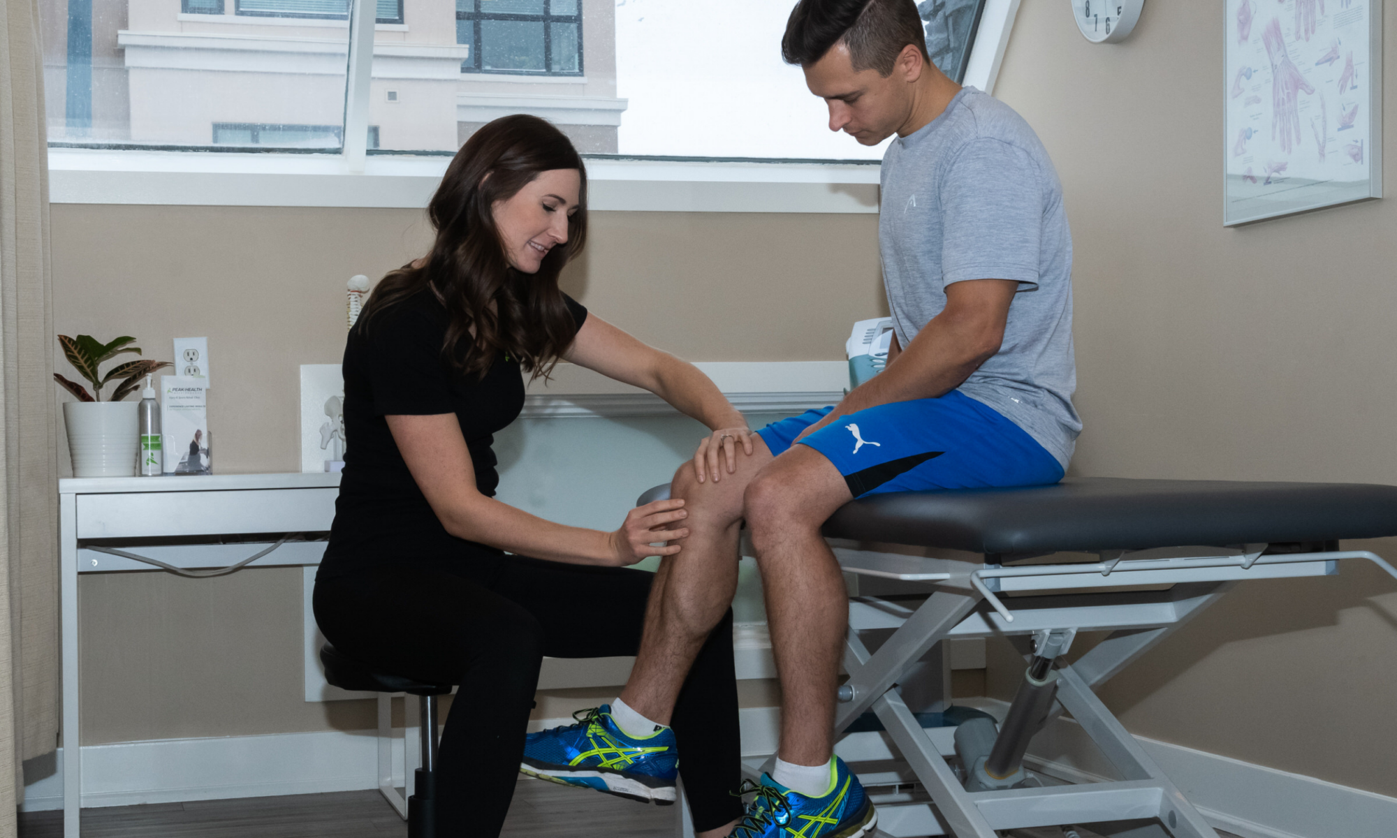
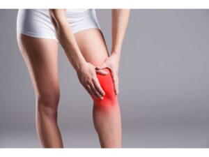
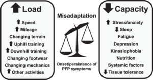
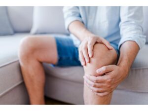
the bus, going up stairs at the office, or squatting to pick up your child. This hits close to home for me, as this was similar to what I was experiencing when  I developed knee pain two months prior to running my first half marathon. With proper activity modification and management of the injury, I was able to complete the half marathon as planned! In this blog, you will learn about the presentation and management of one of the most common types of knee pain, patellofemoral pain syndrome.

What is Patellofemoral Pain Syndrome?

Patellofemoral pain syndrome (aka runner’s knee) is an umbrella term for any long-standing knee pain behind your patella (kneecap) or around the patella. It has also been referred to in literature as runner’s knee or anterior knee pain. Runner’s knee is usually aggravated by any activities performed with your knee in a flexed position, usually while weight bearing. These activities include running, squatting, and stairs. However, your pain may also be aggravated by long periods of sitting. The common theme with all of these is that your knee is undergoing increased compressive forces in a flexed position2,5.

The patellofemoral joint is where your patella (kneecap) articulates with your femur (thigh bone). As you can see in the image above, there are a lot of structures that contribute to the stability and control of the knee and patellofemoral joint. This includes bony structures, ligaments, and muscles. However, stability and control of your knee is also significantly affected by positioning and control of your hip and ankle. So, when you see a physiotherapist, they will likely be addressing any dysfunction at all three of these regions.

Many mechanisms have been proposed as contributing factors to runner’s knee:

      • Patellar maltracking
      • Quadriceps weakness
      • Hip abductor or external rotator weakness
      • Foot pronation
      • Muscular imbalance causing dynamic knee valgus
      • Anatomical abnormalities
      • Overuse or overloading
      • Improper footwear

The literature tends to disagree or be inconclusive on the specific etiology of runner’s knee. Although, a common theme exists in that runner’s knee occurs as a result of biomechanical breakdowns at the hip, knee, and ankle. As a result, this causes a sensitivity in the patellofemoral region, but does not involve any conclusive structural damage.

Why is it Important to Understand Patellofemoral Pain Syndrome?

Patellofemoral Pain Syndrome is one of the most common knee pathologies, 
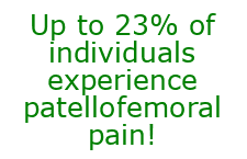
especially for those of you who are runners. It is estimated to have an incidence of 3-15% in active populations and a prevalence of up to 23% in the general population3,4. In my personal caseload, I have treated this condition numerous times, both in adolescents and adults.

When breaking down the stress your knee undergoes when running, it is easy to see why patellofemoral pain is more common in an active population. It is suggested that during running, the ground pushes back on your foot with forces of around 2.5x your bodyweight1. By the time this force is transmitted to the knee, the amount of compression by the quadriceps on the patellofemoral joint can be up to 4x your bodyweight1. This doesn’t mean you shouldn’t be running! This just means that it is important to identify and prevent functional breakdowns in your lower extremity that may lead to injuries associated with the high loads required by running. A physiotherapist or rehab professional will be able to help identify your areas of weakness and work to ensure you can be successful in your sports and leisure activities.

The good news is exercise therapy has strong evidence to support improving short and long term pain in individuals with runner’s knee2. Both strengthening exercises and running retraining have shown to have positive effects in terms of function3. One study suggested education on runner’s knee alone to be more effective than no management at all. The most effective intervention at a 3 month follow up was education combined with physical therapy6.

Taking a step further, recent literature suggests that a multimodal/individualized approach is necessary in treating runner’s knee, as 
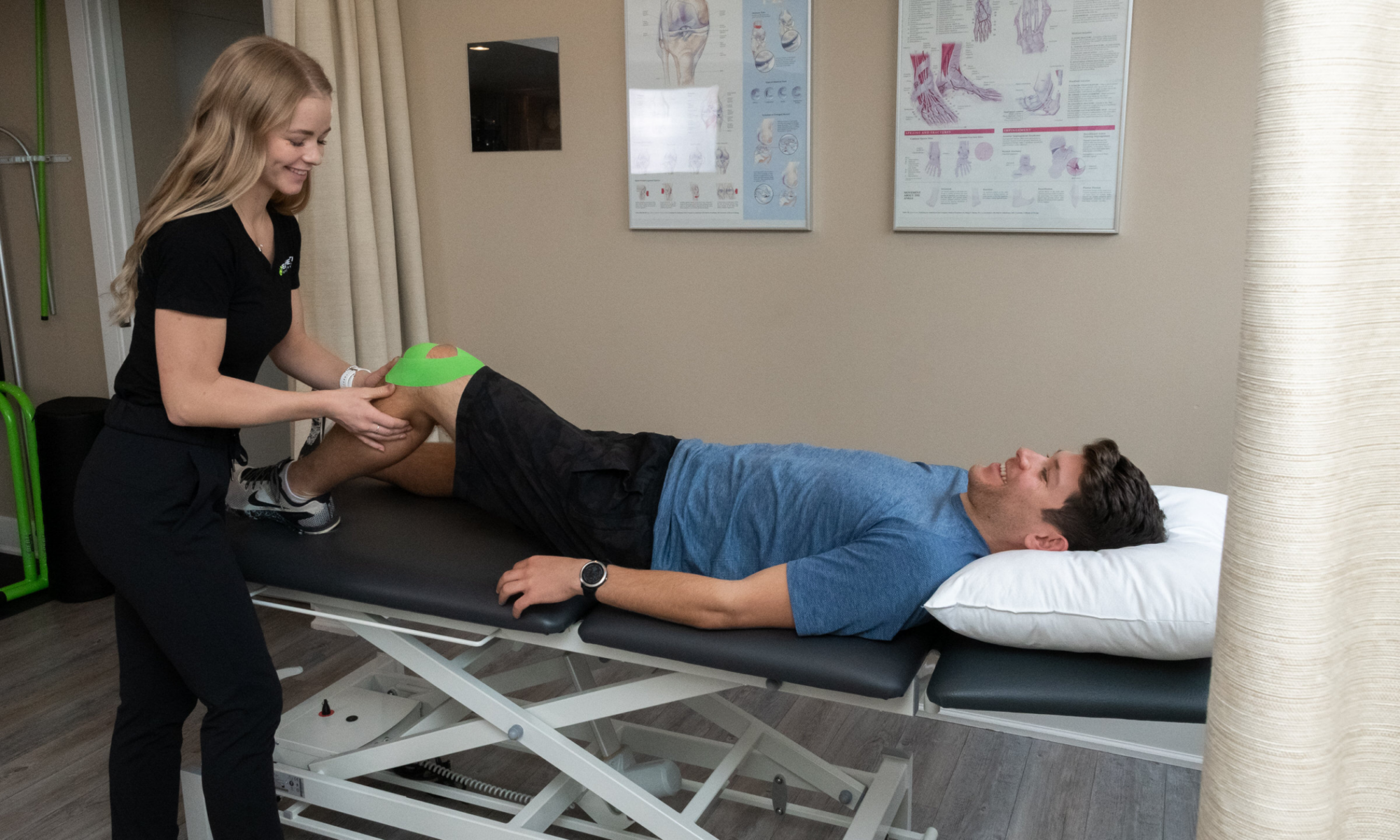
there are many contributing factors to the onset of the pain in the first place1,2. This should include education on taping and footwear.  This should include managing beliefs/expectations on recovery. This should also include education on load/capacity, exercise, and run re-training1. A physiotherapist will be able to support you in most of these areas to give you an opportunity to get back to doing what you love.

Some Risk Factors for Developing Patellofemoral Pain Syndrome2

      • Frequent activity such as running, squatting, and stairs
      • Overuse or sudden increase in physical activity level
      • Quadriceps weakness
      • Dynamic knee valgus (collapsing inwards)
      • Patellar (kneecap) instability
      • Foot abnormalities
      • More common in female sex

*There is a variability and inconsistency in research when it comes to risk factors for developing runner’s knee. Some studies even suggest these issues may be a consequence of patellofemoral pain syndrome, not a cause. This further indicates a need for thorough assessment and multimodal treatment of your knee pain.

How Do I Know If I Have Patellofemoral Pain Syndrome?

      • Knee pain is usually non-traumatic or gradual onset (although you may have a  history of knee trauma/injury).
      • Pain on the anterior (front) of the knee or around the patella (kneecap).
      • You have high activity levels or a recent increase in activity levels.
      • Knee pain with activity such as running, squatting, or stairs.
      • Knee pain with prolonged periods of sitting while your knee is in a flexed/bent position.
      • Knee pain typically does not have associated swelling or locking.

3 Strategies to Help Manage Your Patellofemoral Pain Syndrome Symptoms

1. Activity Modification

*Patellofemoral pain syndrome often occurs as a result of an imbalance between your body’s capacity and the loads it is undergoing with activity. A period of activity modification is likely necessary to manage this imbalance. If you are a runner, try a 1 minute jog, 1 minute walk cycle to see if your pain is better managed.

2. Quadriceps Rolling

*Tight quadriceps can contribute to patellar maltracking related to patellofemoral pain syndrome. Alternatively, quadriceps tightness can come as a result of your knee pain. Rolling will help decrease the tension on the front of your knee when it is in a flexed position. Try 2-5 minutes, 1-2 times per day.

3. Hip Strengthening (Glute Bridge)

*Strengthening the hip girdle will help with biomechanical alignment during dynamic activity such as squatting and running. This will help to decrease your knee pain with activity. Try 3 sets of 10-15 repetitions. You can do this with or without a band around your knees.

FAQ

How long does patellofemoral pain syndrome take to heal?

Healing timelines vary for patellofemoral pain syndrome depending on factors such as your age, activity level, and general health. Pain can often be alleviated with rest and stretching. However, changing the biomechanical breakdowns that contributed to your knee pain takes more time and effort. It is recommended to see a healthcare professional for recommendations to help resolve and prevent your symptoms.

Do I need surgery for my patellofemoral pain syndrome?

Typically, surgery is not required for patellofemoral pain syndrome, as there is no specific structural damage that is causing your pain. If other signs and symptoms exist such as direct trauma to the knee or persistent patellar dislocations, a referral for surgical consultation may be indicated.

What can I not do with patellofemoral pain syndrome?

There are no specific activities that need to be completely avoided with patellofemoral pain syndrome. However, many activities such as squatting, running, and stairs may further aggravate your symptoms. A short term period of rest followed by a gradual increase to full activity is recommended during recovery from patellofemoral pain syndrome.

What Comes Next?

Remember, patellofemoral pain syndrome is knee pain that occurs beneath or around your kneecap. It is one of the most common knee issues, especially for those of you who are active or run a lot. This type of knee pain is typically multifactorial and usually requires individualized management, depending on your specific presentation.

Start by trying some of the strategies listed above and see how it goes! Afterwards, it would benefit you to see a Peak Health physiotherapist to guide you through treatment, depending on your response.

Feel free to reach out if you have any additional questions on runner’s knee or you can book an appointment online by clicking here.

References:

      • Esculier, JF., Maggs, K., Maggs, E., Dubois, B. (2020). “A Contemporary Approach to Patellofemoral Pain in Runners”. Journal of Athletic Training. 55 (12): 1206–1214. https://doi.org/10.4085/1062-6050-0535.19
      • Gaitonde, D.Y., Ericksen, A., Robbins, R.C.; Dwight D. Eisenhower Army Medical Center, Fort Gordon, Georgia (2019). “Patellofemoral Pain Syndrome”. American Family Physician, 99 (2): 88-94, https://www.aafp.org/afp/2019/0115/p88.html?utm_medium=email&utm_source=transaction
      • Neal, B. S., Barton, C. J., Gallie, R., O’Halloran, P., & Morrissey, D. (2016). “Runners with patellofemoral pain have altered biomechanics which targeted interventions can modify: A systematic review and meta-analysis”. Gait & Posture, 45, 69–82. doi:10.1016/j.gaitpost.2015.11.018
      • Neal B.S., Lack S.D., Lankhorst N.E., et al (2019).Risk factors for patellofemoral pain: a systematic review and meta-analysis” British Journal of Sports Medicine; 53: 270-281. https://bjsm.bmj.com/content/53/5/270
      • Physiopedia 2021. “Patellofemoral Pain Syndrome”. Physiopedia. Accessed March 3, 2021, https://www.physio-pedia.com/Patellofemoral_Pain_Syndrome
      • Winters M., Holden S., Lura C.B., et al. (2020). ”Comparative effectiveness of treatments for patellofemoral pain: a living systematic review with network meta-analysis” British Journal of Sports Medicine. doi:10.1136/ bjsports-2020-102819

Media References:

Knee Anatomy Picture:

      • Comprehensive Orthopaedics, S.C. (2016). “Anatomy of the Knee”. Accessed March 3, 2021 via Google Image Search. https://comportho.com/anatomy/anatomy-of-the-knee/. [Original Source Unknown]

Load/Capacity Diagram:

      • Esculier, JF., Maggs, K., Maggs, E., Dubois, B. (2020). “A Contemporary Approach to Patellofemoral Pain in Runners”. Journal of Athletic Training. 55 (12): 1206–1214. https://doi.org/10.4085/1062-6050-0535.19

What is Subacromial Pain Syndrome?

By: Cody Gramlich, Physiotherapist

Shoulder pain can be a frustrating issue, as those of you who have experienced it know. You can’t ignore the ache throughout the day, you can’t 
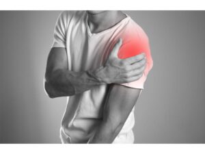
reach to grab a plate from the cupboard, or you can’t roll over at night to sleep on your side. This is what one client was struggling with before choosing to start physiotherapy. With a better understanding of his issue and how to manage it, he was able to return to playing pickleball up to five times per week without shoulder pain! One common cause of shoulder pain is commonly referred to as subacromial impingement, or subacromial pain syndrome. In this blog, you will learn what subacromial pain syndrome is, why it is important to understand, and what you can do to manage it.

What is Subacromial Pain Syndrome?

You may have previously heard the term or been diagnosed with shoulder impingement. However, there has been movement away from the utilization of this diagnosis in recent years, for two main reasons:

    • Impingement only describes a compression mechanism of shoulder pain, whereas subacromial pain syndrome is NOT limited to only a compression of structures.
    • Shoulder impingement as a diagnosis can be further broken down into four categories on its own.

As a result, subacromial pain syndrome is the most up to date terminology used to describe pain that is located on the lateral aspect (outside) or anterior aspect (front) of your shoulder. This type of shoulder pain is non-traumatic and usually worsened with lifting your arm overhead.

Subacromial pain syndrome is an umbrella term that helps describe pain related to any structures in the subacromial space of the shoulder. Your subacromial space is the region located between the acromion of the scapula (bony prominence on the top of your shoulder) and the head of the humerus. This region is displayed in the image below.

Structures included in the subacromial space5:

    • Coracoacromial Arch, composed of the Acromion, Coracoid Process and Coracoacromial Ligaments
    • Humeral Head
    • Subacromial Bursa
    • Tendons of the Rotator Cuff; Supraspinatus, Infraspinatus, Teres Minor and Subscapularis
    • Tendon of the Long Head of Biceps Brachii
    • Coracoacromial ligament
    • G-H Joint Capsule

Since so many structures are located in this region, subacromial pain syndrome encompasses a variety of pathologies that may lead to shoulder pain. This includes conditions such as a shoulder bursitis, biceps tendinopathy, or a rotator cuff issue.

Why Is It Important to Understand Subacromial Pain Syndrome?

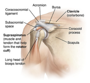
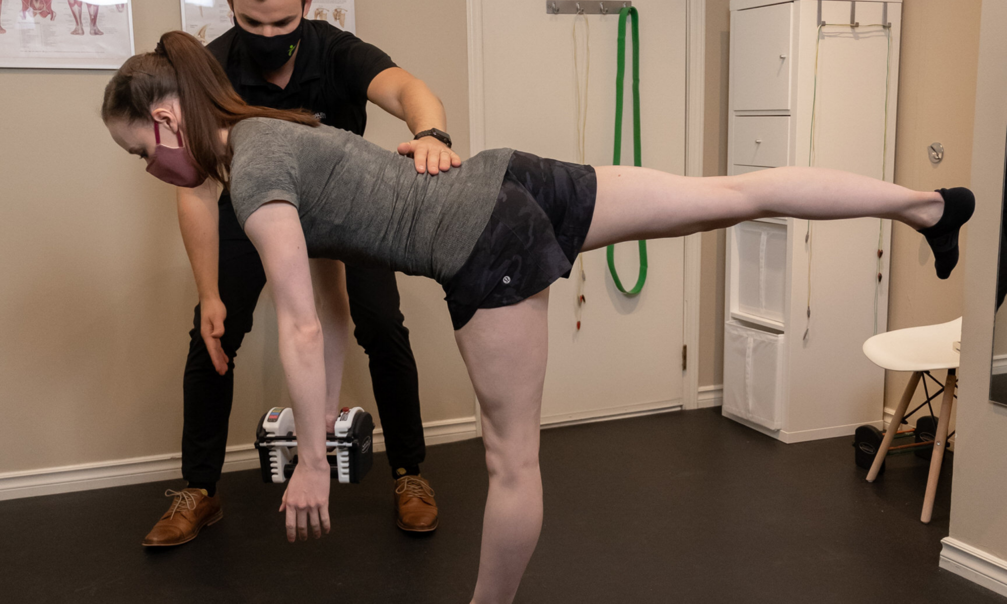
Shoulder pain is common and can result in significant loss of function or participation in your day-to-day activities. It is suggested that up to 67% of community dwelling individuals may experience shoulder pain1. 44-65% of all reports of shoulder pain are thought to involve symptoms arising from the subacromial space1. Subacromial pain syndrome is a prevalent issue, so it is important to understand how to prevent and manage it.

Since subacromial pain syndrome is a generic term that encompasses many structures or more specific diagnoses, it is important that you consult a professional to determine which structures may be most affected. Your treatment can be individualized to your specific presentation. This will lead to optimal outcomes, and help you reach your personal goals.

Regardless of the specific pathology or cause of your subacromial pain syndrome, it has been indicated in many studies that physiotherapy can help. One systematic review made a strong recommendation for exercise therapy as the first-line treatment in subacromial pain syndrome6. A strong recommendation to include manual therapy as an integrated treatment was also made6. Another systematic review even suggested exercise to be as effective as arthroscopic surgery for subacromial pain syndrome3.

Some Risk Factors for Developing Subacromial Pain Syndrome2,5,6

    • Repetitive movements of the shoulder or hand/wrist during work.
    • Work that requires much or prolonged strength of the upper arms.
    • Hand-arm vibration (high vibration and/or prolonged exposure) at work.
    • Working with a poor ergonomic shoulder posture.
    • Altered shoulder kinematics associated with capsular tightness.
    • Rotator cuff and scapular muscle dysfunction.
    • Age (older).

How Do I Know If I Have Subacromial Pain Syndrome?

    • No injury or trauma to cause your shoulder pain (gradual onset)
    • Pain on the anterior (front) or lateral (outer) aspect of your shoulder.
    • Shoulder pain when lifting your arm or with your arm in overhead positions (work or sports with overhead positions).
    • Shoulder pain while lying on your affected side.
    • Shoulder pain when lifting or holding objects in front of your body.

*Remember that these are the most common symptoms that would indicate you may have subacromial pain syndrome. It may present with different signs/symptoms depending on the individual. If this sounds like you, reach out to a physiotherapist or another healthcare practitioner for a thorough assessment to determine the cause of your specific symptoms.

3 Strategies to Help Manage Your Subacromial Pain Syndrome Symptoms

You may have been struggling with shoulder pain recently and are looking for some guidance. As mentioned earlier, research has consistently shown the benefits of physiotherapy. It even suggests supervised exercise should be the first-line management in subacromial pain syndrome.

1) Postural Awareness

*Being aware of your posture will optimize the position of your shoulders and decrease irritation on structures in the subacromial space. Set a timer for every 30-60 minutes to remind yourself to think about your posture or change up your positioning.

2) Thoracic (Upper Back) Mobility

*Decreased upper back mobility or tension in the muscles of the chest can affect posture or shoulder positioning. The exercise above can help with these areas. Try the exercise with your hands across your chest if it is too uncomfortable to have your hands behind your head. Try 2-3 sets of 8-10 repetitions.

3) Rotator Cuff Strengthening

*Strengthening the rotator cuff muscles allows for better positioning and control of the shoulder with activity. This will help lead to decreased shoulder pain. Try 2-3 sets of 8-12 repetitions.

FAQ

How Long Does It Take Subacromial Pain Syndrome to Heal?

Healing timelines vary for subacromial pain syndrome depending on factors such as your age, activity level, and general health. Some of the strongest positive outcomes for subacromial pain syndrome have been shown with a combination manual therapy and supervised exercise program. It is recommended to see a healthcare professional to determine how you can optimize your healing timelines.

Do I Need Surgery for My Subacromial Pain Syndrome?

Conservative treatment and exercise therapy is the first line of treatment for subacromial pain syndrome. It is suggested that conservative treatment should be considered for up to a year and surgery be contemplated only after exhaustive conservative management5. A healthcare professional will be able to help rule out immediate need for surgery and recommend best treatment options for your subacromial pain syndrome.

What Is the Best Way to Sleep With Subacromial Pain Syndrome?

Typically, sleeping on the affected shoulder will be the most uncomfortable position. The ideal sleeping position will change from individual to individual. Sleeping on your back or sleeping on your unaffected side with the painful shoulder supported by a pillow will likely be the most comfortable positions.

What Comes Next?

Remember, subacromial pain syndrome is non-traumatic pain on the front or outside aspect of your shoulder and is typically worsened with lifting your arm overhead. It is thought to be the most common shoulder issue and can significantly affect your ability to participate in hobbies and work. This is why it is important to be able to identify the signs of subacromial pain syndrome and have a plan to treat and prevent the issue.

Start by trying some of the strategies listed above and see how it goes! Afterwards, it would benefit you to see a physiotherapist in Calgary to guide you through treatment depending on your response.

Feel free to reach out if you have any additional questions on shoulder pain or subacromial pain syndrome or you can book an appointment online by clicking here.

References:

    • Chaconas E.J., Kolber M.J., Hanney W.J., Daugherty M.L., Wilson S.H., Sheets C. “SHOULDER EXTERNAL ROTATOR ECCENTRIC TRAINING VERSUS GENERAL SHOULDER EXERCISE FOR SUBACROMIAL PAIN SYNDROME: A RANDOMIZED CONTROLLED TRIAL”. International Journal of Sports Physical Therapy. 12 (7): 1121-1133. (2017) doi:10.26603/ijspt20171121
    • Diercks, R., Bron, C., Dorrestijn, O., Meskers, C., Naber, R., de Ruiter, T., Willems, J., Winters, J., van der Woude, H. J. (2014). “Guideline for diagnosis and treatment of subacromial pain syndrome”. Acta Orthopaedica, 85 (3), 314–322. (2014) doi:10.3109/17453674.2014.920991
    • Haik, M.N., Alburquerque-Sendín, F., Moreira, R.F.C., Pires, E.D., and Camargo, P.R. “Effectiveness of physical therapy treatment of clearly defined subacromial pain: a systematic review of randomised controlled trials”. British Journal of Sports Medicine, 50(18), 1124–1134. (2016) doi:10.1136/bjsports-2015-095771
    • Hanratty, C.E., McVeigh, J.G., Kerr, D.P., Basford, J.R., Finch, M.B., Pendleton, A., and Sim, J. “The Effectiveness of Physiotherapy Exercises in Subacromial Impingement Syndrome: A Systematic Review and Meta-Analysis”. Seminars in Arthritis and Rheumatism, 42 (3), 297–316. (2012) doi:10.1016/j.semarthrit.2012.03.015
    • Physiopedia 2021. “Subacromial Pain Syndrome”. Physiopedia. Accessed February 15, 2021, https://physio-pedia.com/Subacromial_Pain_Syndrome?utm_source=physiopedia&utm_medium=search&utm_campaign=ongoing_internal
    • Pieters, L., Lewis, J., Kuppens, K., Jochems, J., Bruijstens, T., Joossens, L., Struyf, F. “An Update of Systematic Reviews Examining the Effectiveness of Conservative Physical Therapy Interventions for Subacromial Shoulder Pain” Journal of Orthopaedic & Sports Physical Therapy. Volume 50, Issue3, Pages 131-141. (February 2020). https://www.jospt.org/doi/10.2519/jospt.2020.8498

Media References:

Shoulder Anatomy Picture:

    • Fairview Health Services (2019). “Patient Education: The Shoulder Joint”. Accessed February 26, 2021 via Google Image Search. https://www.fairview.org/patient-education/85899. [Original Source Unknown]

What Is Tennis Elbow?

By: Cody Gramlich, Physiotherapist

Tennis Elbow Pain 
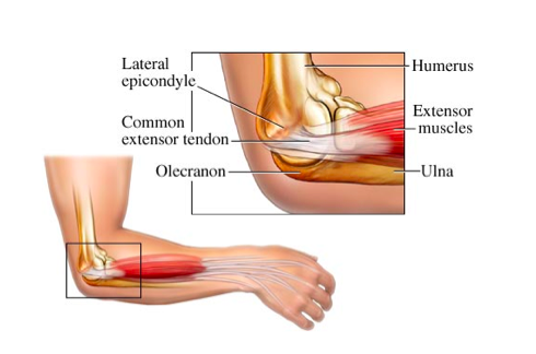
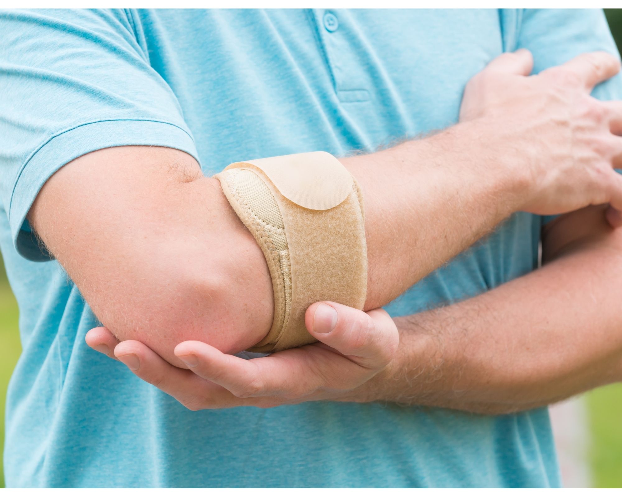
If you have experienced or are experiencing nagging elbow pain, you know that it can significantly affect things such as sleep and your day-to-day activities (think… pouring a cup of coffee, lifting your child, working with hand tools, or playing your favourite sport). This was the story for a client of mine who developed elbow pain after a lot of lifting and carrying during a move. With some guidance and treatment for his issue, he was able to return to skiing weekly, without worrying about elbow pain every time he used his ski poles. If this sounds like something you have been struggling with, or you simply want to learn more, then keep reading! You will learn what tennis elbow is, what to look out for, and how to deal with it.

What Is Tennis Elbow?

Tennis elbow (known as Lateral Epicondylitis) is classified as a repetitive strain or overuse injury. It affects the tendon that attaches to the bony prominence on the outside of your elbow. You typically develop it from activities that involve repetitive, combined wrist and elbow movements. Good examples are golf, racquet sports, and jobs that require frequent tool use.

The wrist extensors are a group of muscles that attach to the bony prominence on the outside of your elbow (lateral epicondyle). These muscles travel down the back of your forearm and across your wrist. They also attach to various points on the back of your hand and fingers. These muscles are primarily involved in extending your wrist and fingers, such as when using a throttle or opening your hand.

What May Cause Tennis Elbow?

Tennis elbow occurs as a result of overuse of your wrist extensor muscle group. This overuse leads to changes in your common extensor tendon structure that will lead to pain on the outside of your elbow.

Tennis is not the only activity that can cause this condition, as it can be any activity that has repetitive combined wrist and elbow movement. Some examples include:

  • Jobs that involve a lot of gripping, such as a painter
  • A month of home renovations
  • Playing guitar around the campfire with your friends

Why Is It Important to Understand Tennis Elbow?

It is suggested in multiple studies that tennis elbow affects 1-3% of individuals2,4,5. My current caseload includes 5-10% of patients needing physical rehab for it!

The statistics above may even underestimate the actual number of individuals suffering from tennis elbow. Many of the studies do not account for those who do not seek care for the condition. There is also a lack of data from worker’s compensation claims, which likely make up a portion of incidents2.

Tennis elbow can significantly affect your ability to work and participate in the activities you enjoy. One specific study that monitored the condition in participants for over 13 years between 2000 and 2012 indicated that 16% of those with tennis elbow had work restrictions. Of those participants, 4% had to take anywhere between 1-12 weeks off of work. Tennis elbow can have a huge impact on your day-to-day life, which is why it is important to identify the condition early and seek the help of a physiotherapist in Calgary. This same study recorded a recurrence rate of 8.5% for the condition.

Proper Tennis Elbow Physiotherapy Treatments

A lot of you may be involved in activities that put you at risk for developing tennis elbow. This can be your occupation, your leisure activity/hobby, or your competitive sport.

Some Risk Factors for Developing Tennis Elbow1,4:

  • Handling tools heavier than 1 kg
  • Handling loads heavier than 20 kg at least 10 times per day
  • Repetitive movements for more than 2 hours per day
  • Age 30-50
  • Smoking
  • Poor tennis mechanics
  • Oral corticosteroid use
  • Comorbid conditions such as DeQuervain’s, Carpal Tunnel Syndrome, or Rotator Cuff Pathology

How Do I Know if I Have Tennis Elbow?

  1. Pain on the lateral (outside) aspect of your elbow.
  2. Typically your pain comes on gradually and worsens with time, but can come on after a single incident of heavy activity.
  3. Radiating pain/ache into the back of your forearm or hand.
  4. Activity related pain on the outside of your elbow with wrist and hand movements..
  5. Weakness of your wrist/hand or decreased grip strength with lifting/carrying.

*Remember that these are the most common symptoms that would indicate you may have tennis elbow. It may present with different signs/symptoms depending on the individual. If this sounds like you, reach out to a health care practitioner for a thorough assessment to determine the cause of your specific symptoms.

3 Strategies to Help Manage Your Symptoms

You may be experiencing some of these issues but know that it is not all bad. One study showed the long-term success rate for a group being treated with physiotherapy to be 91%3. Physiotherapy can help!

1. Use a Tennis Elbow Strap

*Using a tennis elbow strap can be a short-term solution to help manage your pain during activities. It creates a different fulcrum for your extensors, reducing load/tension on the common extensor tendon.

2. Wrist Extensor Rolling

*Rolling the extensor muscles can help resolve any muscle tension associated with tennis elbow and help with your pain. Try rolling the muscles on the back of your forearm for 1-5 min with moderate to deep pressure.

3. Wrist Extensor Strengthening

*Adequately loading or strengthening your common extensor tendon will encourage positive tissue changes and tissue remodeling. Try three sets of 8-10 repetitions, slow and controlled.

FAQ

What is the Best Treatment?

There are variable treatment methods for tennis elbow including injections (cortisone, PRP), prescription management, physiotherapy, and surgical management. Conservative management such as physiotherapy and chiropractic are usually the first line of treatments. You should see a healthcare professional to help determine which method of treatment would be best for you.

Can Tennis Elbow Heal on Its Own?

Tennis elbow can last anywhere between two weeks to two years, with variable recovery rates if left untreated. Treatment is recommended to help manage symptoms, improve recovery timelines, and prevent recurrence.

What Happens if Tennis Elbow Goes Untreated?

Generally, tennis elbow will not lead to serious problems if it goes untreated. However, you may suffer from continued symptoms and further negative structural changes to the tendons in the elbow. This could lead to further injury or a chronic issue and result in missed work and decreased activity participation.

What Comes Next?

Remember, tennis elbow presents as pain on the outside of your elbow as a result of activities that include repetitive gripping or combined wrist and elbow movements. It is a common issue and can significantly affect your ability to participate in hobbies and work. This is why it is important to be able to identify the signs for the condition and have a plan to treat and prevent the issue.

Start by trying some of the strategies listed above and see how you respond. Afterwards, it would benefit you to see a rehabilitation professional to guide you through treatment depending on your response.

Feel free to reach out if you have any additional questions on tennis elbow or you can book an appointment online by clicking here.

References:

  1. Physiopedia 2021. “Lateral Epicondylitis”. Physiopedia. Accessed January 25, 2021, https://www.physio-pedia.com/Lateral_Epicondylitis#cite_ref-53
  2. Sanders, Thomas L., Kremers, Hilal Maradit, Bryan, Andrew J., Ransom, Jeanine E., Smith, Jay, and Morrey, Bernard F., “The Epidemiology and Health Care Burden of Tennis Elbow. A Population-Based Study”. The American Journal of Sports Medicine. 43, no. 5 (February 2015). https://doi.org/10.1177/0363546514568087
  3. Smidt, Nynke, van der Windt, Daniëlle AWM, Assendelft, Willem JJ, Devillé, Walter LJM, Korthals-de Bos, Ingeborg BC, Bouter, Lex M. “Corticosteroid injections, physiotherapy, or a wait-and-see policy for lateral epicondylitis: a randomised controlled trial”. The Lancet, 359 (2002). https://www.physio-pedia.com/images/9/9e/Smidt_et_al_RCT_lateral_elbow.pdf
  4. Springer Science+Business Media New York. Winston, Jonathan and Wolf, Jennifer M. “Chapter 1. Tennis Elbow: Definition, Causes, Epidemiology”. Tennis Elbow, 1, no 6 (2015). doi:10.1007/978-1-4899-7534-8_1
  5. Vicens, Gemma, Seijas, Roberto, Sallent, Andrea, Dominguez, Andrea, Ares, Oscar, and Torrecilla, Andrea. “Tennis Elbow Pathogenesis”. International Journal of Orthopaedics. 4, no. 3 (June 2017). http://96.126.98.199/index.php/ijo/article/view/1988/2416

Media References:

Elbow Anatomy Picture:

  1. Mountain Health and Performance. “Tennis Elbow Treatment North Vancouver”. Accessed February 5, 2021 via Google Image Search. http://www.mountainhp.ca/conditions-treated/what-is-tennis-elbow/. [Original Source Unknown]

Foot Pain Exercises for Runners

Dr. Logan from Peak Health and Performance takes time in this video to show you some simple exercises that you can do to overcome foot pain while running. He will show you the technique that you need to do in order to make them effective, and He will talk about how often you need to do the exercises in order to get optimal results. Dr. Logan is a chiropractor at Peak Health that focuses on sports injuries as well as chronic injuries in the active population. If you suffer from foot pain while running or cycling, or just get foot pain in general, this video is for you!

Protect Your Hamstrings with Dr. Riley Sjodin

Dr. Riley Sjodin of Peak health and Performance shows three ways to help strengthen and add capacity to your hamstrings so that you can run and partake in your summer activities pain-free and injury free. These three exercises are simple and easy to do and require very little equipment making them easy to do at home. If you have any questions regarding these exercises please do not hesitate to contact Dr. Riley at our clinic or seek the care or advice from any other member of our sports injury team.

The Clamshell Exercise for Glute Activation & Lower Limb Stability

Janelle Juss, PT, discusses the importance of doing the clamshell exercise properly. This exercise can be an excellent first step in knee and hip rehab to make sure there is ample glute activation to stabilize the lower limb. Keeping the hips still during the movement and not using momentum is key! Let us know if you have any questions or if you would like the next step in lower limb stability and rehabilitation.

Peak is a safe, welcoming, and inclusive place. Be yourself, we like it that way.